

Veterinary Ultrasound Services in Lahore & Karachi


Non-Invasive Imaging for a Deeper Look
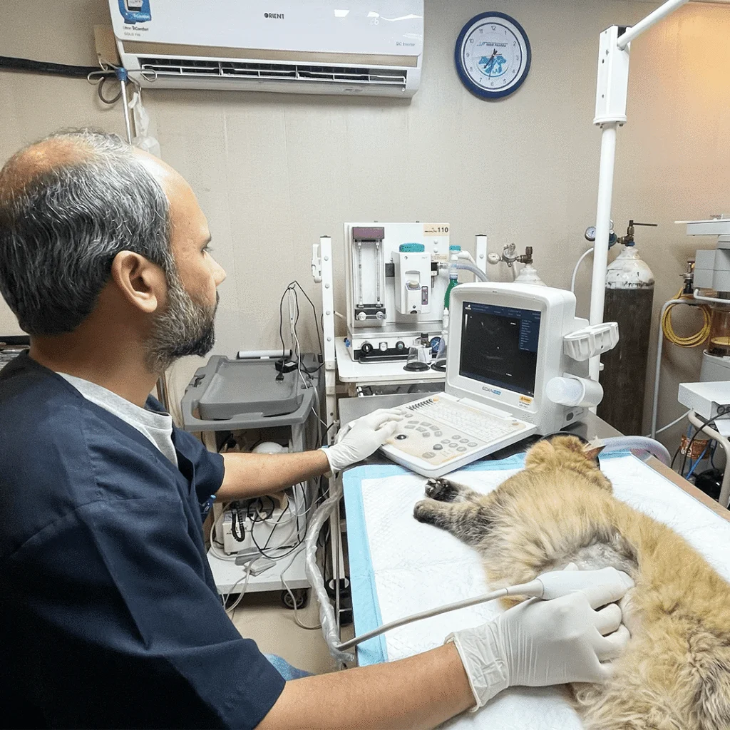
Ultrasound uses sound waves to create real-time images of internal organs, blood flow, and developing pregnancies. Unlike X-rays, ultrasound provides exceptional soft tissue detail — invaluable for abdominal organs, the heart, and reproductive structures.
Our ultrasonography is performed by trained professionals using high-resolution equipment for detailed abdominal, cardiac, and musculoskeletal imaging.
Common Uses of Veterinary Ultrasound
Abdominal ultrasound evaluates liver, kidneys, spleen, bladder, adrenals, pancreas, and GI tract to detect tumours, cysts, organ enlargement, fluid, and stones. Cardiac ultrasound (echocardiography) assesses heart structure, valve function, and blood flow for heart disease diagnosis. Pregnancy confirmation provides early detection, litter size estimation, and foetal
viability monitoring. Ultrasound-guided biopsy and aspiration enables precise sampling of masses for lab analysis.
Ultrasound is painless and typically needs no sedation. A small area may be shaved for image quality. Your pet lies comfortably while gel is applied and the probe is passed over the area. The procedure takes fifteen to thirty minutes.
DHA Karachi
- 61-C , Zulfiqar Commercial Avenue , Phase - VIII , DHA Karachi, Pakistan.
- +923139999759
- VIEW ON MAP


Frequently Asked Questions
For abdominal ultrasound, we recommend fasting eight to twelve hours as a full stomach can obscure other organs. Water is fine.
No. It is completely pain-free and most pets tolerate it calmly without sedation.
They are complementary. X-rays are better for bones and chest; ultrasound excels at soft tissue. Your vet will recommend the appropriate modality.
Most ultrasound examinations take between 20 to 40 minutes, depending on the area being assessed.